Radiographie und Tomographie mit Spaltneutronen
wissenschaftlicher Leiter: Adrian Losko
Das Radiographie- und Tomographiesystem NECTAR an der Forschungs-Neutronenquelle Heinz Maier-Leibnitz (FRM II) der Technischen Universität München wurde von RCM konzipiert, instrumentiert und seit Inbetriebnahme des FRM II bis ca. 2020 von der RCM betreut. Aktuell erfolgt die Betreuung durch den Instrumentverantwortlichen Adrian Losko vom FRM II. Das weltweit einzigartige Instrument nutzt schnelle Neutronen aus der Kernspaltung des 235U zur Durchleuchtung unterschiedlichster Objekte und Materialien.
Weitere Informationen finden sich unter https://mlz-garching.de/medapp-nectar/de.
Visualization of water uptake in a trunk
Dr. Thomas Bücherl, Dr. Christoph Lierse von Gostomski
Introduction
The visualization of dynamic processes in sealed objects is of vital interest for many investigations and applications. For example, it results in knowledge on the mechanisms of root growing in soil allowing the optimization of watering and manuring for plant growing. The visualization of oil distributions in running or starting engines can give information on further aspects for its optimization reducing the fuel consumption. Both examples, like many more, might have large economic and ecological impacts.
The main aspect of non-destructive inspection techniques should be that the samples investigated are in their natural surroundings. Plants should be placed in large sized soil containers, i.e. wall effects should not influence their growing, engines should not require special windows to be inserted to get access to the required information, i.e. the processes visualized should not be influenced by using different materials etc. Thus, often large and / or dense samples have to be investigated. Here, radiography using fission neutrons can give valuable information. These neutrons have a high penetration power for dense materials while being sensitive to hydrogen containing materials like water, oil etc.
A highly interesting material for these types of investigations is wood, as it is of manifold use even in sensitive areas like building construction, and especially its reactions with water. As an example the water uptake in a trunk was investigated.
Experiment
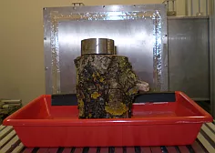
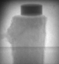
NECTAR at FRM II is a versatile facility for radiography, tomography and activation measurements using fission neutrons [1]. The neutron spectrum can be adapted to the specific requirements of the individual experiments by means of different filters and collimators. For the real time measurements of a trunk of about 12 cm diameter a neutron flux of 5.4×105 cm-2s-1 and a L/D of about 230 were selected. The trunk was placed in an empty bowl and loaded by an iron cylinder on the top to avoid its floating when water was filled in the bowl (Fig. 1). Using a CCD based detection system, operated at a temperature of -50° C, first a radiography of the initial set-up (i.e. trunk in bowl without water) was measured (Fig. 2). This was referred to as the reference image. Then, the bowl was filled with about 250 ml of water and a series of 1000 radiographies back-to-back was started immediately, each lasting 60 seconds, thus covering a time period of about 52 hours.

Fig. 1. Photography of the experimental set up for the measurement of water uptake in a trunk. The trunk is loaded by an iron cylinder. In the background the converter plate of the detector system is visible

Fig. 2. Radiography of the initial set-up. The image of the trunk shows the structures of the bark and of the inner area, both. The horizontal line in the lower part of the image corresponds to the upper rim of the bowl. On top of the trunk, the iron cylinder is visible.
Results
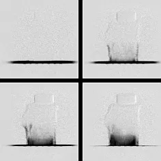
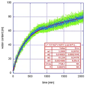
All images were filtered [2], dark image corrected, normalized and then subtracted by the adequately treated reference image. Fig. 3 shows some of these difference images for 2 minutes, 200 minutes, 1000 minutes and 2000 minutes after the filling of the water in the bowl, respectively. The dark areas indicate the location of water, i.e. after 2 minutes nearly all water is still in bowl while after 2000 minutes the water is mainly in lower part of the trunk. A qualitative investigation showed that within the first 200 minutes the water is mainly soaring within the bark until it reaches a maximum height of about 5 cm. Here gravitation seems to prevent a further ascent, i.e. is leveling out the effect of the transport mechanism in the bark. For the remaining time the water uptake in the inner parts of the trunk is the dominant effect while the amount of water in the bark decreases. These results are confirmed by a quantitative data evaluation [3]. The soaring within the bark, well described by an exponential rise, is the dominant mechanism for the first 750 minutes, while for the remaining 1250 minutes, the water uptake in the inner areas of the trunk becomes dominant and is described by a linear function (Fig. 4). At the end of the experiment about 80 ml of water are absorbed by the trunk.

Fig. 3. Radiographies of the water uptake in a trunk at different time intervals after adding water in the bowl. Upper left: After 2 minutes only the water in the bowl is visible. Upper right: After 200 minutes soaring within the bark has started. Lower left: After 1000 minutes the uptake within the trunk started. Lower right: After 2000 minutes the water content in the bark is reduced

Fig. 4 Results of the quantitative evaluation of the experiment (Blue: water content derived from radiograph in millilitre; green: corresponding uncertainty; red: fit on data)
Conclusion
It is shown that real time measurements of slow processes on bulky objects like a trunk are possible at the NECTAR facility using fission neutrons. The visualization of the dynamics of such processes can act as a starting point for new or improved physical models trying to describe reality more accurate. Although in this experiment the time frame was quite large (60 seconds), it can be reduced to a few seconds or even lower but actually on the expense of spatial resolution.
References
[1] T. Bücherl, et al., Nucl. Instrum. Methods A, to be published (2011).
[2] K. Osterloh et all., Nucl. Instrum. Methods A, to be published (2011).
[3] T. Bücherl, et al., Nucl. Instrum. Methods A, to be published (2011).